Importance of Testing for Heart Disease
Approximately 80 million Americans have some form of heart disease. Saint Francis Medical Center’s Heart Hospital uses the latest tests and procedures to diagnose heart-related disorders before they become fatal. Stress tests, blood pressure monitoring, and angiography are effective diagnostic tools.
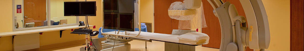
Angiography
An angiography is an X-ray test used to detect diseases of the blood vessels, such as the weakening of the vessel walls and the narrowing or blocking of vessels. The X-ray is taken after the vessels are injected with a substance that enables them to be captured on film.
Blood pressure Monitoring
Because high blood pressure is one of the three main factors associated with heart attacks, it is important to check your blood pressure regularly. We monitor the blood pressure of our Heart Hospital patients around the clock.
CT
The computerized tomography (CT) scanner helps identify aortic disease, cardiac masses and pericardial diseases.
64-slice CT
The 64-slice CT is a powerful computer-driven scanner that can capture vividly detailed 3-D images in 5-10 seconds (compared to 30-40 seconds for previous CT scanners) and provide unprecedented clarity with full-color, high-resolution imaging for early detection of various diseases and pathologies to more accurately diagnose and treat patients.
Cardiac Calcium Scoring
Cardiac calcium scoring uses high-speed CT technology to take rapid pictures of the heart and the arteries around it to detect calcium or plaque buildup. Plaque causes hardening of the arteries and can lead to heart attack if left untreated. A scan is assigned a score from 0 (no calcification) to 400 (significant calcium buildup).
EKG
An electrocardiogram (EKG) measures the heart’s electrical activity to help diagnose abnormal heart conditions. The physician monitors the EKG readings to check for irregularities in the heart rhythm.
Fractional Flow Reserve
This measurement process occurs during the heart catheterization to aid in detecting a blockage significant enough to need intervention.
Holter Monitoring
During a heart Holter monitor study, a patient wears a monitor (usually for 24 hours) that records electrical activity of the heart. Saint Francis also offers 30-day Holter monitoring for appropriate cases. While being monitored, the patient also records a diary of his/her activity. Heart care specialists then analyze the data to diagnose any irregularities.
Intravascular Ultrasound
Intravascular ultrasound enables doctors to view coronary anatomy during diagnostic and interventional catheterization to identify a blockage that is significant enough to require intervention.
MRI
Magnetic resonance imaging (MRI) uses powerful magnets to look inside the body. Computer-generated pictures can show the heart muscle, identify damage from a heart attack, diagnose certain congenital cardiovascular defects and evaluate disease of larger blood vessels.
P.E.T. Scan
A positron emission tomography (P.E.T.) scanner is an imaging test that shows how organs function rather than just how they look. Functional abnormalities usually occur long before structural damages are evident, making early detection of disease possible.
Stress Tests
- Exercise Stress Test: Physical exercise is performed on a treadmill while your heart is monitored. If you are having nuclear images done with this exam, a radioactive medication is administered while you are exercising.
- Chemical Stress Test: If you are unable to perform physical exercise, then your physician may order a chemical stress test. Various chemicals including Adenosine, Dobutamine, Persantine or Lexiscan may be used to exercise the heart in place of physical exercise. If you are having nuclear images done with this exam, a radioactive medication is administered as well. This type of stress test is always done with either an echocardiogram or nuclear imaging.
- Stress Echocardiogram: In a stress echocardiogram, ultrasound images of your heart are taken during various stages of cardiac stress. Depending on your physical condition, you may have an exercise stress test or chemical stress test associated with your stress echocardiogram.
- Nuclear Stress Test: You will have two injections of a radioactive medication to allow for images of the blood flow to your heart. One injection and image will before the stress test and one injection will be during the stress test with images after. Depending on your physical condition, you will have either an exercise or chemical stress test as listed above associated with the imaging.
Transesophageal echocardiogram
A transesophageal echocardiogram is a special type of imaging procedure that uses a tube with a transducer on the end of it. The tube is passed down your throat into the esophagus to get very clear images of your heart and its structure.